
RESEARCH GROUP – MINERALOGY
Team:
 |
 |
 |
 |
 |
| ANDREOZZI Giovanni | BALLIRANO Paolo | BOSI Ferdinando | PACELLA Alessandro | MACRI' Michele |
Topics:
The GEO/06 sector carries out research activities on two main topics: Environmental Mineralogy and Crystal Chemistry/Crystal physics of Minerals.
Environmental mineralogy
|
|
|
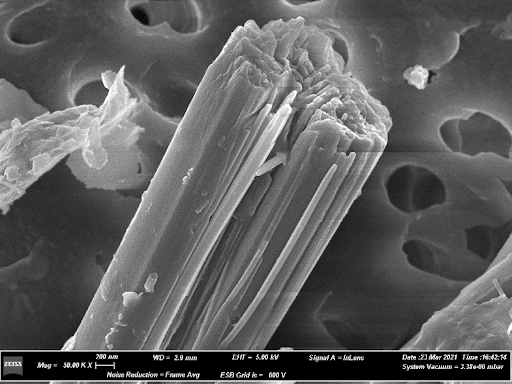
| TEM bright-field images of (a) pristine tremolite fibers and (b) thermally treated at 1200 °C. In (c) two pyroxene crystals associated with amorphous material; (d) detail showing pyroxene grains associated with amorphous material (taken from J. Haz. Mat. 2020, 398, 123119). | FE-SEM image of erionite fibres |
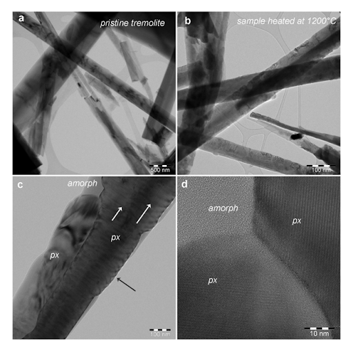
| We perform the structural and crystal chemical characterization of asbestos and other mineral fibres not yet restricted or regulated. We study the modifications induced, both at the bulk and surface, by their prolonged immersion in solutions mimicking the body fluids to unravel correlations between the physical chemical features of the mineral fibres and their toxicity/carcinogenicity. We are evaluating the hazard of the so-called NOA (Naturally Occurring Asbestos), i.e. fibrous minerals in their natural setting. We investigate the relationships between lithology, mineralogy and structural setting of asbestos bearing outcrops and the crystal chemistry of the fibres. We perform thermal treatments of ACM (Asbestos Containing Materials) aimed at their inertisation by analysing in situ the ongoing structural, chemical and morphological modifications induced by heating and we evaluate the residual reactivity of the fibres. These topics are truly interdisciplinary and are studied using a multi-analytical approach. The results are published in highly ranked journals. |
 |
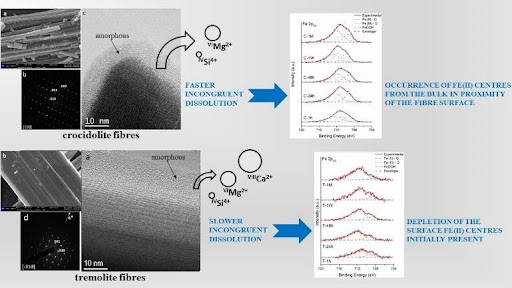 |
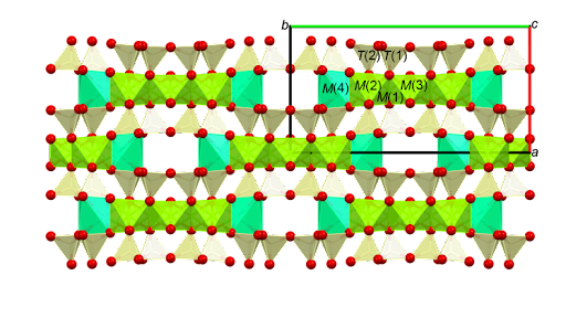
| Projection as seen along the c-axis of the structure of tremolite (taken from Mineral Mag. 2020, 84, 888). | Sketch showing the different kinetics of dissolution of tremolite and crocidolite in mimicked body fluids. |
Mineral crystal chemistry and crystal physics
|
|
|
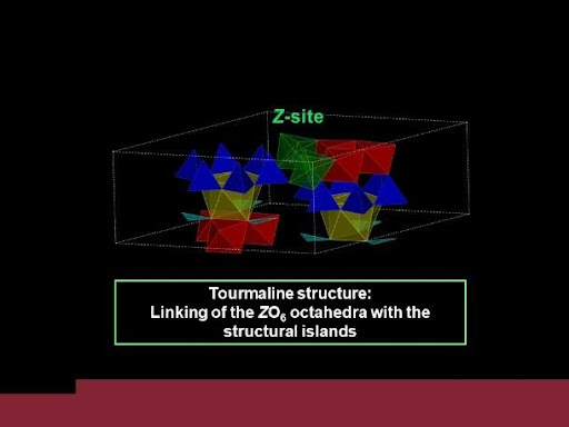
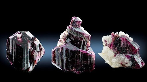
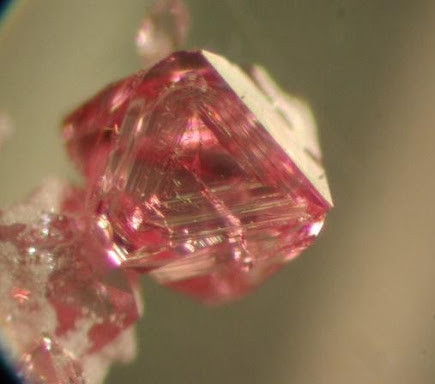
| Representation of the tourmaline structure with the structural islands, made of X (yellow), Y (red), B (cyan) and T (blue) polyhedral, connected by the Z octahedral (green). | Crystals of Mn-bearing purplish-red tourmaline from Madagascar, up to 4 cm in size (photo by Federico Picciani) |
| The objective of our research is the definition of genetic conditions, crystal chemistry, physical properties and HP-HT behavior of minerals. We clarified the general crystal chemistry of minerals belonging to the spinel, tourmaline and axinite supergroups, and we are among the leaders at international level for the study of these minerals. We are now investigating their thermodynamic stability, modifications as a function of P-T-X conditions, breakdown conditions and products, and their behavior in different geologic environments, from granitic pegmatites to subduction zones. Measuring physical properties of these minerals has several implications and applications in Earth and planetary Sciences, Materials Science and in Gemology. Furthermore, our group is actively involved in finding new minerals and defining mineral classification criteria and nomenclature rules. This activity led us to discover dozens of new minerals and to define the nomenclature and classification of important mineral groups. |
 |
 |
| Octahedral spinel crystal, flux growth synthesis (about 0.6 mm, optical microscope picture, 100x) |
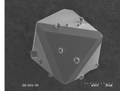
Octahedral spinel crystal, flux growth synthesis (about 0.06 mm, SEM picture) |